INTRODUCTION
Microorganisms need nutrients, a source of energy and certain environmental conditions in order to grow and reproduce. In the environment, microbes have potential to adapt to the habitats most suitable for their needs, but then it is hard for microbes to do so in the laboratory. This is when a growth media or also known as culture media plays an important part. This is basically an aqueous solution to which all the necessary nutrients have been added, depending on the type and combination of nutrients, different categories of media can be made. A growth medium or culture medium is a liquid or gel designed to support the growth of microorganisms or cells. Different types of cells prefers different type of media, depending on their needs. There are actually two major types of culture media, one of it are those used for cell culture, which use specific cell types derived from plants or animals. The second one is microbiological culture, which are used for growing microorganisms, such as bacteria or yeast. The most common growth media for microorganisms are nutrient broths and agar plates.
The composition of self-made agar broth is listed below :
1.5 g/L “Lab-lemco” powder (a beef extract)
1.5 g/L Yeast extract
5.0 g/L Peptone (a nitrogen source)
5.0 g/L Sodium chloride
15.0 g/L Agar powder
For your information, the self-made agar broth actually contains the same composition with the manually-made nutrient medium, except that it contains 15 g/L agar. We must also ensure that the final pH value of both medias is 7.4 .
Speaking of autoclaving, autoclaves are more or less like pressure cookers very similar to the ones that you see in the stores. As we know, food cook a lot faster in a pressure cooker than they do in a regular pot or in the oven. This is due to the intense heat and pressure that is applied to the food. The same mechanism works against living microorganisms in an autoclave. Once an autoclave is started, steam is pushed into the chamber that contains the items that are being sterilized. As the steam goes in, the pressure and temperature within the chamber is increased. Most autoclaves are set to increase steam pressure until a temperature of at least 121 degrees Celsius is reached. This temperature and pressure will remain at this level for at least 15 minutes. This is a high enough temperature for a long enough period of time to kill any and all microorganisms and their spores. The steam and pressure are released and brought down to normal room temperature and pressure after the 15 or more minutes of running.
 |
| Image 1 : Example of culture media in agar plates |
OBJECTIVE
To prepare sterile nutrient agar for culturing microorganisms .
MATERIALS AND REAGENTS
Commercial Nutrient Agar
Brain Heart Infusion Broth ( BHI )
Trypticase Soy Broth ( TSAYE )
Peptone powder
Beef extract powder
Sodium chloride
Yeast extract
Electronic Weighing Balance
Distilled water
Scott bottles
Measuring cylinder
Glass rod
Beakers
PROCEDURE
A . Commercial nutrient agar
1 . 11.2 g of the commercial nutrient agar is weighted using an electronic
weighing balance and placed into a beaker.
2 . 400 ml of distilled water is measured using a measuring cylinder and poured into
the beaker containing the nutrient agar. The solution is then stirred by using a
glass rod until it mixes well.
3 . After the solution is mixed well, the solution is poured into the Scott bottle that
had been sterilized.
4 . The bottle is the loosely recapped and is set aside and ready to undergo
sterilization in an autoclave machine.
5 . The media is sterilized at 121 degree Celsius for 15 minutes.
6 . The media is removed after 15 minutes of autoclaving. The media is allowed to
cool down and the cap of the bottle is tighten.
B . Self-made nutrient agar
1 . 0.60 g of beef extract, 0.6 g of yeast extract, 2.0 g of peptone, 2.0 g of sodium
chloride and 6.0 g of agar powder are weighed using an electronic weighing
balance and placed into a beaker.
2 . 400 ml of distilled water is measured using a measuring cylinder and poured into
the beaker containing the nutrient agar. The solution is then stirred by using a
glass rod until it mixes well.
3 . After the solution is mixed well, the solution is poured into the Scott bottle that
had been sterilized.
4 . The bottle is the loosely recapped and is set aside and ready to undergo
sterilization in an autoclave machine.
5 . The media is sterilized at 121 degree Celsius for 15 minutes.
6 . The media is removed after 15 minutes of autoclaving. The media is allowed to
cool down and the cap of the bottle is tighten.
C . Brain Heart Infusion agar (BHI)
1 . 5.20 g of BHI agar in powder form is weighed using an electronic weighing
balance and placed into a beaker.
2 . 100 ml of distilled water is measured using a measuring cylinder and poured into
the beaker containing the nutrient agar. The solution is then stirred by using a
glass rod until it mixes well.
3 . After the solution is mixed well, the solution is poured into the Scott bottle that
had been sterilized.
4 . The bottle is the loosely recapped and is set aside and ready to undergo
sterilization in an autoclave machine.
5 . The media is sterilized at 121 degree Celsius for 15 minutes.
6 . The media is removed after 15 minutes of autoclaving. The media is allowed to
cool down and the cap of the bottle is tighten.
D . Trypticase Soy Agar ( TSAYE )
1 . 4.00 g of TSAYE agar in powder form is weighed using an electronic weighing
balance and placed into a beaker.
2 . 100 ml of distilled water is measured using a measuring cylinder and poured into
the beaker containing the nutrient agar. The solution is then stirred by using a
glass rod until it mixes well.
3 . After the solution is mixed well, the solution is poured into the Scott bottle that
had been sterilized.
4 . The bottle is the loosely recapped and is set aside and ready to undergo
sterilization in an autoclave machine.
5 . The media is sterilized at 121 degree Celsius for 15 minutes.
6 . The media is removed after 15 minutes of autoclaving. The media is allowed to
cool down and the cap of the bottle is tighten.
RESULTS
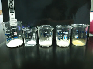
4 different culture media was prepared which are 400 ml of commercial nutrient agar, 400 ml of self-made nutrient agar, 100 ml of Brain Heart Infusion (BHI) agar and 100 ml of Trypticase Soy Agar ( TSAYE ). The composition of the materials needed are stated in the procedure. They are weighed approximately and dissolved with distilled water. Stirring of the solution takes place until the solution is dissolved and mixed. After mixing, it is only poured into sterilized Scott bottles and ready to place into an autoclave machine for 15 minutes at the temperature of 121 degree Celsius.
 |
| Image 2 : Weighting the appropriate amount using an electronic weighting balance |
 |
| Figure 3 : Done weighing the 6.00 g agar powder for self-made nutrient agar |
 |
| Figure 4 : The preparation for self-made nutrient agar |
 |
| Figure 5 : Appropriate amount of distilled water obtained using a measuring cylinder |
 |
| Figure 6 : Transfer of the solution after mixing with distilled water into Scott bottles |
 |
| Figure 7 : Four different cultured media prepared |
 |
| Figure 8 : Cultured media ready to undergo autoclaving |
 |
| Figure 9 : Cultured media in an autoclave |
DISCUSSION
1 . There are actually a few precautions that we need to take note throughout the
experiment, which is :
- The pan of the electronic weighing balance is cleaned with a small brush first to prevent any small leftover particles that might affect the weight reading.
- The “tare” button is pressed every time after the empty beaker is put on the balance to obtain accurate measurements and to prevent zero errors.
- When using a measuring cylinder to obtain distilled water, make sure that the position of the eye is at the same level as the bottom of the meniscus ( surface of water that is curved downwards ) to prevent parallax errors.
- All of the apparatus used are cleaned and rinsed using distilled water before using.
- The media is stirred well using a glass rod to ensure balance mixing and to maintain the concentration of the media.
- Make sure that the caps of the Scott bottles are only slightly tightened to prevent the Scott bottles from breaking during autoclaving.
2 . Before the Scott bottles with different medium are placed into the autoclaving machine for sterilization, there are some steps that need to be followed as below :
- The drain screen at the bottom of the chamber is checked before using the autoclave.
- Any debris noticed is cleaned up for efficient heat transfer as steam must flush out of the autoclave chamber. If the drain screen is blocked with debris, a layer of air may form at the bottom of the autoclave and prevent proper operation.
- The water level is ensured to be higher than the bottles in the autoclave.
- The cover of the autoclave chamber and exhaust valve is tightened.
- The temperature is checked so it is always maintained at 121°C and the pressure is ensured to reach 103 kPa above the atmospheric pressure, with steam is continuously forced into the chamber.
- The time for destruction of the most resistant bacterial spore is now reduced to about 15 minutes. For denser objects, up to 30 minutes of exposure may be required. The conditions must be carefully controlled or serious problems may occur.
- Then, the exhaust valve is opened to ensure the pressure drops to nearly 0 kPa before removing the basket with Scott bottles from the autoclave chamber.
CONCLUSION
As a conclusion, we are able to learn the correct steps and methods to prepare different media for culturing microorganisms. Precautions must also remembered when carrying out the steps and procedures. Preparation and sterilization of culture media is important to prevent contamination of the unwanted microorganisms inside the media. We also obtained the information that autoclaving is actually a fast and efficient sterilization process.
Reference
2 . Obtained from
http://www.sigmaaldrich.com/analytical-chromatography/microbiology/learning-center/theory/media-preparation.html on 24 October 2015
3. Obtained from
October 2015
4. Obtained from https://en.wikipedia.org/wiki/Autoclave on 24 October 2015


