2.1 Ocular Micrometer
INTRODUCTION
An ocular micrometer is a glass disk that fits in a microscope eyepiece that has a ruled scale, which is used to measure the size of magnified objects. In other words , it serves as a scale or ruler to measure the size of magnified objects. The physical length of the marks on the scale depends on the degree of magnification. The ruler on a typical ocular micrometer has between 50 to 100 individual marks, is 2 mm long and has a distance of 0.01 mm between marks. Technicians can easily make calculations of object size after measuring an object against the dimensions of an ocular micrometer, which are calibrated using a stage micrometer, a microscope slide with its own surface scale visible when viewed through the microscope . The function of the stage micrometer is to achieve calibration of the microscope by checking the divisions and measurements on the eyepiece reticle. The distance between the lines of an ocular micrometer is an arbitrary measurement that only has meaning if the ocular micrometer is calibrated for the objective being used. In the world of science, an ocular micrometer is usually used to measure the sizes of prokaryotes and eukaryotes. It is also used to categorized newly found microorganisms and distinguish them based on their sizes .
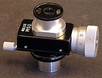 |
| Figure 1 : Micrometer eyepiece |
 |
| Figure 2 : An Ocular micrometer |
OBJECTIVE
1 . To learn the correct steps and procedures of using an ocular micrometer.
2 . To measure and count cells using a microscope
MATERIALS AND REAGENTS
Microscope fitted with an ocular micrometer , slide micrometer and stained preparation.
PROCEDURE
( Refer to manual )
RESULTS
The ratio of magnification is calculated by using the formula below,
One ocular division = No. of division on stage micrometer
No. of division on ocular micrometer
When using 10 x objective lens , 1 mm on stage represent 9.6 ocular divisions
When using 40 x objective lens , 0.1 mm on stage represent 8.0 ocular divisions
For 40 x objective lens ,
The length of cells : 8.0 ocular unit = 0.1 mm
0.2 ocular unit = 2.50 µm
The width of cells : 8.0 ocular unit = 0.1 mm
0.1 ocular unit =1.25 µm
 |
| Figure 3 : 40 x objective lens , 400 maginification |
 |
| Figure 4 |
DISCUSSION
1 . We have to ensure that the first line of the ocular micrometer is in line with the
first line of the stage micrometer to avoid errors when reading the measurements,
this is what be call calibration .
2 . In this experiment, parallax errors should be prevented when calibrating the
ocular micrometer with the stage micrometer to get an accurate calibration.
3 . An ocular micrometer does not have any units, therefore it is a must for it to be
calibrate with the stage micrometer to get the correct unit and scale.
REFERENCE
2 . Obtained from http://www.medilexicon.com/medicaldictionary.php?t=55222 on the
the 10th of October 2015
2.2 Neubauer Chamber
Neubauer chamber also known as a hemocytometer is a device used to count cells and was originally designed for counting red blood cells. Touching a little on the history of the hemocytomer, it was actually invented by Louis-Charles Malassez. The Neubauer chamber is a thick crystal slide with the size of a glass slide of 30 x 70 mm and 4 mm thickness . A neubauer chamber consists of a thick glass microscope slide with a rectangular indentation that creates a chamber. This chamber is engraved with a laser-etched grid of perpendicular lines. The device is carefully crafted so that the area bounded by the lines and depth is known. It is therefore possible to count the number of cellsor particles in a specific volume of fluid, and thereby calculate the concentration of cells in the fluid overall. In a simple counting chamber, the central area is where cell counts are performed. The chamber has three parts. The central part, where the counting grid has been set on the glass. Double chambers are most common than simple chamber. In this case, the chamber has two counting areas than can be loaded mm in size. The grid has 9 square subdivisions of width 1 mm. Cover slips for counting chambers are specially made thicker than those for conventional microscopy, since they must be heavy enough to overcome surface tension of a drop of liquid. The cover slip is placed over the counting surface prior to putting on the cell suspension.
 |
| Figure 5 : A nerbauer chamber |
OBJECTIVE
1 . To practice how to use a neubauer chamber precisely to count the cell .
MATERIALS AND REAGENTS
Cell line culture, neubauer chamber and cover slip, Sterile Pastuer pipettes .
PROCEDURE
( Refer to manual )
RESULTS
Total number of cells in 9 random small squares = 5+6+5+5+6+5+6+4+4
= 46
The average number of cells per square = 46 / 9
= 5.11 cells
Volume of the square = 0.25 mm x 0.25 mm x 0.1 mm
= 6.25 x 10⁻³ mm 3
= 6.25 x 10⁻⁶ cm
= 6.25 x 10⁻⁶ mL
This shows that there are 5.11 cells in 6.25 x 10⁻⁶ mL , therefore the concentration of cells = 5.11 cells / 6.25 x 10 -6 mL
= 8.176 x 10 5 cells / mL
 |
| Figure 6 : Image of cell line on hemacytometer |
DISCUSSION
1 . A few drops of cell line culture is transfered to the surface of the hemocytometer
by using aseptic techniques practiced in the previous lab. The specially made
cover slip is a must to be placed on the hemocytometer to give a precise volume
in the space delimited by the grid and the cover slip.
2 . The cover slip must be placed on the surface of hemocytometer carefully to avoid
the air bubbles from forming .This is because the air bubbles actually block our
vision when examining the cell in the counting chamber. Cover slip for counting
chambers is specially made and thicker than those for conventional microscopy.
Since they must be heavy enough to overcome the surface tension of a drop of
liquid.
3 . 9 small squares are selected randomly without being picky from a total of 25
square to get an accurate result . If any of the cells are located between two
boxes , it should be counted only once but not twice . It is also advisable not to
count cells which are further expose outside the square .
4 . Then at last, the average number of cells is obtained and the concentration of
cells can be calculated .
CONCLUSION
With the help of an ocular micrometer, we are able to measure magnified specimens or microorganisms under the microscope. On the other hand, with the help of a Neubauer chamber or hemocytometer, we are able to calculate the cell concentration in a culture and get a rough number of cells in the culture.
REFERENCE
1 . Obtained from http://www.celeromics.com/en/resources/Technical%20Notes/cover-cell-glass.php on 10th of October 2015
No comments:
Post a Comment