1.1 Setting up and using the microscope
Introduction :
A microscope is an optical instrument that enables us to see the objects which are not visible to the human naked eye. The most common type of microscope that we usually use in the lab is a compound light microscope. This microscope has two lenses that bend light so that a specimen is magnified and projected. A light microscope without a doubt uses visible light, which is the part of the light spectrum our eyes can see, and passes it through the lenses. The lenses bend the light so that the object which is being observe appears larger than it is, which allows us to see better.
Parts of the light microscope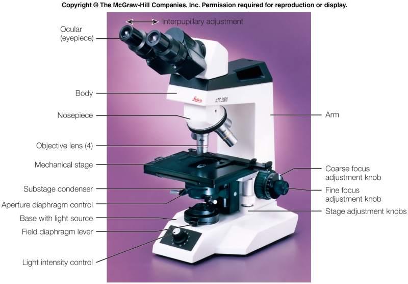
The light microscope, the most common type of microscope, contains several parts with specific functions. The light is the main source of illumination which comes from the base of the instrument. From the top, the eye piece lens is where you can place your eyes to look through the microscope . It contains the ocular lens, which usually provides a standard magnification power of 10x. The tube connects the eyepiece to the objective lenses. The nosepiece holds the objective lenses and can be rotated easily to change magnification.
There are 4 objective lenses on a microscope. They almost always consist of 4X, 10X, 40X and 100X powers. When coupled with a 10X (most common) eyepiece lens, we get total magnifications of 40X (4X times 10X), 100X , 400X and 1000X. The shortest lens is the lowest power, the longest one is the lens with the greatest power. The nosepiece can be adjust so that you can turn from one objective to the other, depending on how much magnification you need for your specimen.
The mechanical stage acts as a platform where the slides is placed. To hold the slides in place, stage clips can be used. The slides are flexible and can be moved around while it is on the stage by using the stage adjustment knob.
Coarse adjustment knob is the large, round knob on the side of the microscope used for focusing the specimen. It may move either the stage or the upper part of the microscope. On the other hand, the fine adjustment knob is a small, round knob on the side of the microscope used to fine-tune the focus of your specimen after using the coarse adjustment knob.
If you notice carefully, there is a hole in the stage which is called aperture, that allows light through for better viewing of the specimen. The diaphragm controls the amount of light going through the aperture. And lastly, the light or mirror usually found near the base of the microscope acts as the source of light.
MAGNIFICATION AND RESOLUTION
Magnification is the ability to make small objects seem larger, such as making a microscopic organism visible. The total magnification of the image can be calculated by using the formula which is, Objective lens multiplication X Eyepiece lens multiplication.
Therefore, four magnifications will be produced by the microscope:
4x objective X 10x eyepiece = 40x magnification
10x objective X 10x eyepiece = 100x magnification
40x objective X 10x eyepiece =400x magnification
100x objective X 10x eyepiece =1000x magnification
Resolution is the ability to distinguish two objects that are very close together from each other. The term magnification is often confused with the term resolution. While high magnification without high resolution may make very small microbes visible, it will not allow the observer to distinguish between microbes or sub-cellular parts of a microbe. To be able to distinguish between two objects under a microscope, a viewer must first magnify to a point at which resolution becomes relevant . Resolution is affected by adjusting the condenser diaphragm. Closing the diaphragm increases the contrast of the image but decreases the resolution ; while by opening the diaphragm, contrast is decreased but resolution is increased.
Magnification and resolution are equally important to produce a clear image while using the microscope. One without another will not be enough to produce a good result. For an example, if the magnification used is high but the resolution is low , only a fuzzy image will be produced and vice versa.
OBJECTIVE
Learn how to use a simple bright-field microscope correctly
MATERIALS AND REAGENTS
Microscope slide and cover-slip
PROCEDURE
( Refer to manual )
RESULTS
 |
| V. cholerae under 40x magnification |
 |
| V. cholerae under 100x magnification |
 |
| V. cholerae under 400x magnification |
 |
| V. cholerae under 1000x magnification |
Discussion
After being able to observe the ‘X’ on the marker pen slide , we are also able to observe a specimen slide called V. cholerae by using 40x, 100x and 400x magnification. Vibrio cholerae is a Gram-negative, comma-shaped bacterium. Some strains of V. cholerae cause the disease cholera. V. cholerae is a facultative anaerobic organism and has a flagellum at one cell pole.
Scientific classification of V . Cholerae :
Domain : Bacteria
Phylum : Proteobacteria
Class : Gammaproteobacteria
Family : Vibrionaceae
Genus : Vibrio
Morphology :
Shape : curved or comma-shaped
Colour : Red
REFERENCE
2. Obtained from http://sciencelearn.org.nz/Contexts/Exploring-with-Microscopes/Science-Ideas-and-Concepts/Magnification-and-resolution on 2nd of October 2015
3. Obtained from http://utahscience.oremjr.alpine.k12.ut.us/sciber00/7th/cells/sciber/micrpart.htm on 2nd of October 2015
4. Obtained from http://microbiologyonlineblog.blogspot.my/2009/11/microbiology-online-focus-on-parts-and.html on the 2nd of October 2015
1.2 Examination of the cell
INTRODUCTION
Bacteria which is under the Kingdom Prokaryote are very minute organisms. Because of it’s size that is too small, bacteria are seldom studied under low-power or high-power objective lens. Instead, they are stained first and observed with the oil immersion objective. Aseptic technique can be used during the transfer of microorganisms. Aseptic technique is a method designed to prevent contamination from microorganisms. It involves applying the strictest rules and utilizing what is known about infection prevention to minimize the risks that you’ll experience an infection.
A wet mount is one of the most common ways of observing specimens under the microscope. The water or oil performs an important optical function. Without it, the resolution is lower. Therefore, in order to study the bacteria in its natural state, we have to use wet mount technique. The wet mount technique enables us to study the sizes , shapes of living microorganisms and to determine whether or not the cells are motile.
OBJECTIVE
OBJECTIVE
a) To provide an experience in the use of microscope
b) To illustrate the diversity of cells and microorganisms
MATERIALS AND REAGENTS
Lactobacillus sp. culture
Immersion oil
Lens tissue
Inoculating loop
Bunsen burner
Slide and coverslip
Immersion oil
Lens tissue
Inoculating loop
Bunsen burner
Slide and coverslip
PROCEDURE
( Refer to manual )
( Refer to manual )
RESULTS
DISCUSSION
 |
| Lactobacillus sp. observed under a light microscope |
Lactobacillus ( genus lactobacillus), any group of rod-shape, gram-positive, non-spore forming bacteria of the family Lactobacillaceae. Lactobacillus is a member of lactic acid bacteria group . ( its members convert lactose and other sugars to lactic acid). Lactobacillus are generally non-motile and can survive in both aerobic and anaerobic environments. Examples of other Lactobacillus species include L.acidophilus, L.brevis, L. Casei and L. Sanfranciscensis.
They are usually straight, although they can form spiral or coccobacillary forms under certain conditions. They are often found in pairs or chains of varying length. Lactobacillus are classified as lactic acid bacteria, and derive almost all of their energy from the conversion of glucose to lactate during homolactic fermentation. In this process 85-90% of the sugar utilized is converted to lactic acid. They generate ATP by nonoxidative substrate-level phosphorylation.
Lactobacilli produce lactic acid and are used for many different things, including yogurt production and the maintenance of healthy intestinal microflora. Lactobacilli are commonly associated with the gastrointestinal tract of humans. The genome of the Lactobacillus plantarum has been sequenced and the genomes of several other Lactobacilli are underway.
CONCLUSION
In conclusion, by the end of the experiment, we learned the techniques and steps of how to use a bright field microscope and ways to take good care of the microscope. Other than that, the steps of aseptic technique was also practiced. It is important to practice aseptic technique to prevent contamination. From what we observed from the microscope, we understand that by using oil immersion technique with high power objective lens of 100x which results in the magnification of x1000, the microorganism , Lactobacillus can be seen clearer. The Lactobacillus was prepared with wet mount observer, therefore we can see the bacteria in its natural state without needing any staining procedures.
REFERENCE
1 . Obtained from http://www.healthline.com/health/aseptic-technique#Overview1 on 4th of October 2015
2 . Obtained from https://microbewiki.kenyon.edu/index.php/Lactobacillus on the 4th of October 2015
3 . Obtained from http://www.microbehunter.com/making-a-wet-mount-for-microscopy/ on the 4th of October 2015
4 .Obtained from http://global.britannica.com/science/Lactobacillus on the 4th of October 2015
I have become happy to have a look at this newsletter after searching at google, after reading I have written a chunk of the article about apple nutrition chart Thank you for the thing and supporting me.
ReplyDelete